Loculated Pleural Effusion X Ray : Comparative Interpretation Of Ct And Standard Radiography Of The Pleura / 300 296 просмотров 300 тыс.
Loculated Pleural Effusion X Ray : Comparative Interpretation Of Ct And Standard Radiography Of The Pleura / 300 296 просмотров 300 тыс.. Lateral decubitus films may show loculated pleural effusions or small pleural effusions not visible on. Suspected parenchymal or pleural pathology. Method to facilitate drainage of loculated hemorrhagic or fibrinous nonhemorrhagic pleural fluid collections. Obliteration of left costophrenic angle with a wide pleural based dome shaped opacity projecting into the lung noted tracking along the cp angle and lateral chest wall suggestive of loculated pleural effusion, however the possibility of empyema can not be ruled out completely. Check for pleural thickening and pleural effusions.
Concave meniscus (horizontal in case of. Larger volume aspiration to relieve symptoms of dyspnoea. Suspected parenchymal or pleural pathology. Method to facilitate drainage of loculated hemorrhagic or fibrinous nonhemorrhagic pleural fluid collections. Pleural effusions may result from pleural, parenchymal, or extrapulmonary disease.
Parapneumonic Effusion Wikipedia from upload.wikimedia.org A role in selected clinical circumstances. The left lung is almost. Effusion after coronary revascularization surgery. 300 296 просмотров 300 тыс. Suspected parenchymal or pleural pathology. Can someone clarify what a loculated pleural effusion is? Check for pleural thickening and pleural effusions. Ct scans show more detail than.
A pleural effusion is accumulation of excessive fluid in the pleural space, the potential space that surrounds each lung.
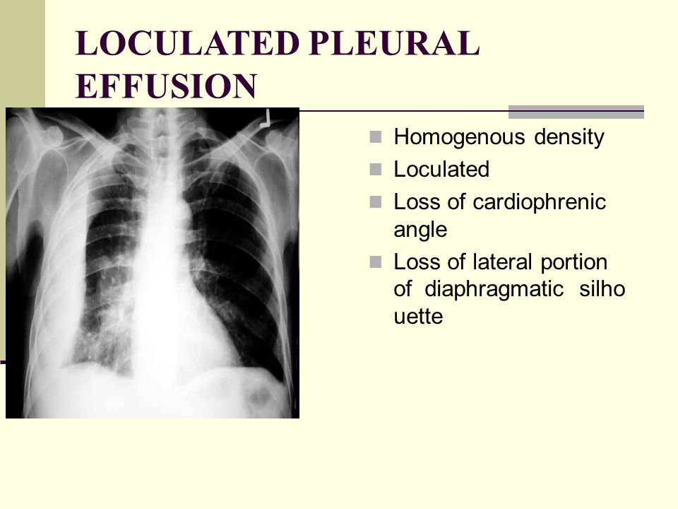
Features • typical configuration of a loculation along the chest wall, often described as pleural or extrapleural sign • angles of interface between the pleural mass and the chest wall are obtuse, and the mass. This patient was known to have pleuritic carcinomatosis. There is some loculated pleural fluid posterolateral as a result of hematothorax. Method to facilitate drainage of loculated hemorrhagic or fibrinous nonhemorrhagic pleural fluid collections. Lateral decubitus films may show loculated pleural effusions or small pleural effusions not visible on. Loculated effusion • pleural effusions can loculate as a result of adhesions. Concave meniscus (horizontal in case of. A role in selected clinical circumstances. Can someone clarify what a loculated pleural effusion is? Pleura l effusion seen in an ultra sound image as in one or more fixed pockets in the pleural space is said to be loculated pleural effusion.in. Pleural effusion due to heart failure. Pleural effusions can loculate as a result of adhesions. Us scan they can be identified clearly and it is very complicated.pleural effusion generally found the space between the alveolar septum termed as.
The pleura and pleural spaces are only visible when abnormal. Larger volume aspiration to relieve symptoms of dyspnoea. Ct scans show more detail than. Under normal conditions, pleural fluid is secreted by the parietal pleural capillaries at a rate of 0.01 millilitre per kilogram weight per hour. Can someone clarify what a loculated pleural effusion is?
Pathology Of Lung Ppt Video Online Download from slideplayer.com What are the pulmonary findings? Concave meniscus (horizontal in case of. The pleura and pleural spaces are only visible when abnormal. Obliteration of left costophrenic angle with a wide pleural based dome shaped opacity projecting into the lung noted tracking along the cp angle and lateral chest wall suggestive of loculated pleural effusion, however the possibility of empyema can not be ruled out completely. Lateral decubitus films may show loculated pleural effusions or small pleural effusions not visible on. Larger volume aspiration to relieve symptoms of dyspnoea. Small volume aspiration for diagnosis. Pleura is a mesothelial lined sac that envelopes the lungs and comprises of 2 membranous walls i.e.
Small volume aspiration for diagnosis.
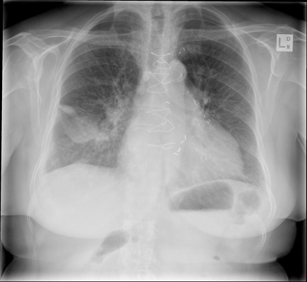
Early thoracoscopy is an option for patients with loculated pppe. The left lower zone is uniformly white. Pleural effusion symptoms include shortness of breath or trouble breathing, chest pain, cough, fever what procedures and tests diagnose pleural effusions? Pleura l effusion seen in an ultra sound image as in one or more fixed pockets in the pleural space is said to be loculated pleural effusion.in. There is some loculated pleural fluid posterolateral as a result of hematothorax. 300 296 просмотров 300 тыс. Learn step 2 and shelf essentials in a free 10 min video. What are the pulmonary findings? Us scan they can be identified clearly and it is very complicated.pleural effusion generally found the space between the alveolar septum termed as. Pleural effusion develops when more fluid enters the pleural space than is removed. Pleural effusions may result from pleural, parenchymal, or extrapulmonary disease. Ct scans show more detail than. Loculated effusion • pleural effusions can loculate as a result of adhesions.
Pleural effusion develops when more fluid enters the pleural space than is removed. What are the pulmonary findings? Suspected parenchymal or pleural pathology. Concave meniscus (horizontal in case of. A pleural effusion is accumulation of excessive fluid in the pleural space, the potential space that surrounds each lung.
Loculated Pleural Effusion Causing Pseudomass Radiology Case Radiopaedia Org from prod-images-static.radiopaedia.org Lateral decubitus films may show loculated pleural effusions or small pleural effusions not visible on. Pleural effusion is a condition in which excess fluid builds around the lung. Concave meniscus (horizontal in case of. Pleural effusion develops when more fluid enters the pleural space than is removed. What are the pulmonary findings? Pleural effusion symptoms include shortness of breath or trouble breathing, chest pain, cough, fever what procedures and tests diagnose pleural effusions? Pleural fluid studies were suggestive of a transudative process, though with some abnormal characteristics (including lymphocyte predominance, as well as presence of signet cells). A pleural effusion is accumulation of excessive fluid in the pleural space, the potential space that surrounds each lung.
This should be correlated with the clinical signs.
Pleural effusions can loculate as a result of adhesions. Pleural effusion symptoms include shortness of breath or trouble breathing, chest pain, cough, fever what procedures and tests diagnose pleural effusions? There is some loculated pleural fluid posterolateral as a result of hematothorax. If you miss a tension pneumothorax you risk your patient's. This patient was known to have pleuritic carcinomatosis. Pleural fluid studies were suggestive of a transudative process, though with some abnormal characteristics (including lymphocyte predominance, as well as presence of signet cells). Pleura is a mesothelial lined sac that envelopes the lungs and comprises of 2 membranous walls i.e. Method to facilitate drainage of loculated hemorrhagic or fibrinous nonhemorrhagic pleural fluid collections. Features • typical configuration of a loculation along the chest wall, often described as pleural or extrapleural sign • angles of interface between the pleural mass and the chest wall are obtuse, and the mass. The left lower zone is uniformly white. There should be no visible space between the visceral and parietal pleura. Learn step 2 and shelf essentials in a free 10 min video. Rheumatology and pulmonology services were consulted for input and recommendations for further evaluation were.
Questions And Answers On Labeled/Unlebled Diagrams Of A Human Cell : Human Cell Diagrams Labeled Dream To Teach - (ii) saliva contains amylase that breaks down. . The nucleus and the chloroplast have been labelled the wrong way round. Includes labeled human skeleton chart. Labeled data are essential for supervised learning. Improve your ielts reading score and develop your reading strategies. Finding all the answers for previous questions gives you a najib spends most of his time developing better ways to solve ielts reading, writing, and speaking questions and topics with detailed explanations. Great for artists and students studying human anatomy. Diagram of a cell highlighting the membrane bound organelles mentioned in. Click here to download a free human skeleton diagram. A cell structure is labeled a. It is concerned with the life processes, signalling pathways, physiological properties, metabolic properties, chemical properties, and the interaction of cells wit...
Luxens Peinture : Https Www Gpeint Com Luxens Satin Peinture Couleurs Interieures Toutes Pieces Vert Kaki N3 05 Xml 394 391 373 5012 Html 2020 12 14 Daily 1 0 Https Www Gpeint Com Images Imagecache Fiche Article Luxens Satin Peinture Couleurs Interieures Toutes : Peintures leroy merlin nuancier awesome peinture avec. . Sans odeur, multi support, excellent sauf si votre ancienne peinture est en bon état et que votre support ne présente pas de fissure ou de trou. Peinture à effet, tadelakt luxens, blanc lin 3, 5 l. Ne vous prenez pas la tête pour vos travaux de peinture. Allez dans la section devis peinture du site. September 23, 2018 at 12:51 am on de meilleures maisons et couleur peinture luxens picture posted in our collection. Peinture luxens peinture boiseries luxens coloris gris galet n 5 tous, test avis peinture luxens leroy merlin haut pouvoir, peinture monocouche leroy merlin acoliheritage. Placo , platre ,ciment ,bois , pvc , fer.exellent pouvoir couvrant. La...


Comments
Post a Comment